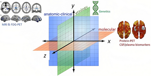
Atrophy in the medial temporal lobe (MTA) is being used as a criterion to support a diagnosis of Alzheimer’s disease (AD). There are several structural neuroimaging approaches for quantifying MTA, including semiquantitative visual rating scales, volumetry (3D), planimetry (2D), and linear measures (1D). Current applications of structural neuroimaging in Alzheimer’s disease clinical trials (ADCTs) incorporate it as a tool for improving the selection of subjects for enrollment or for stratification, for tracking disease progression, or providing evidence of target engagement for new therapeutic agents. It may also be used as a surrogate marker, providing evidence of disease-modifying effects. However, despite the widespread use of volumetric magnetic resonance imaging (MRI) in ADCTs, there are some important challenges and limitations, such as difficulties in the interpretation of results, limitations in translating results into clinical practice, and reproducibility issues, among others. Solutions to these issues may arise from other methodologies that are able to link the results of volumetric MRI from trials with conventional MRIs performed in routine clinical practice (linear or planimetric methods). Also of potential benefit are automated volumetry, using indices for comparing the relative rate of atrophy of different regions instead of absolute rates of atrophy, and combining structural neuroimaging with other biomarkers. In this review, authors present the existing structural neuroimaging approaches for MTA quantification. They then discuss solutions to the limitations of the different techniques as well as the current challenges of the field. Finally, and due to their relevance, they discuss how the current advances in AD neuroimaging can help AD diagnosis.
Reference: Journal of Alzheimer's Disease, DOI: 10.3233/JAD-150226 Full text
Reference: Journal of Alzheimer's Disease, DOI: 10.3233/JAD-150226 Full text
Comparison of volumetry, planimetry, linear measures, and visual scales in the assessment of medial temporal lobe atrophy on MRI.Reproducibility is considered very good when ICC >0.90), good when ICC is between 0.80–0.90, moderate 0.60–0.80 and poor
| Visual rating Scales | Linear methods (1D) | Planimetric methods (2D) | Volumetric methods (3D) | |
| Objective and reliable | No | Yes | Yes | Yes |
| Requirements of | Any MRI scan | Any MRI scan | Any MRI scan | May need specific |
| MRI studies | is suitable | is suitable | is suitable | MRI studies |
| Time consuming | Little | Little | Little | Extensive |
| Training | Little | Little | Little | Extensive |
| Software | Not required | Any software for MRI | Any software for MRI visualization is suitable | Special software for volumetric studies |
| Reproducibility | Poor | Still needs to be tested | Good | -Automated: good to very good |
| - Manual: depends on experience | ||||
| Feasible in clinical practice | Yes | Yes | Yes | Not widely available |
| Feasible in clinical trials | Still needs to be tested | Still needs to be tested | Still needs to be tested | Yes |


